Perforated Peptic Ulcer-A Silent Killer in Gut
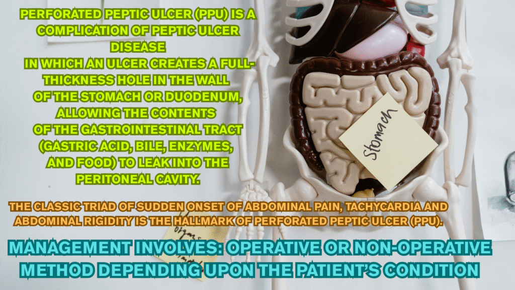
Perforated peptic ulcer (PPU) is a complication of peptic ulcer disease in which an ulcer creates a full-thickness hole in the wall of the stomach or duodenum, allowing the contents of the gastrointestinal tract (gastric acid, bile, enzymes, and food) to leak into the peritoneal cavity.
The classic triad of sudden onset of abdominal pain, tachycardia and abdominal rigidity is the hallmark of perforated peptic ulcer (PPU).
Risk Factors of Perforated peptic ulcer:
- Excessive use of NSAIDs
- Helicobacter pylori( pylori)
- physiological stress
- smoking
- corticosteroids
- previous history of PUD (Peptic ulcer disease).
Pathogenesis of PPU:
Continuous damage to the mucosal lining of stomach and duodenum leads to the formation of such perforation.
H.pylori infections and the use of NSAIDs and low-dose aspirin are known to damage the mucosal lining.
In the case of NSAID (and aspirin) use, mucosal damage is secondary to inhibition of cyclooxygenase 1 (COX-1) derived prostaglandins which play crucial role in maintaining mucosal integrity. Once the mucosal layer is disrupted, the gastric epithelium is exposed to acid, and the ulcerative process begins. If the process continues, the ulcer deepens reaching the serosal layer. A perforation occurs once the serosal layer is ruptured at which point the gastric contents are released into the abdominal cavity.
Symptoms of PUD (peptic ulcer disease) include:
Symptoms of peptic ulcer disease when left untreated leads to the development of Perforated peptic ulcer. So, it’s very essential to know first about the symptoms of peptic ulcer disease. Here are the most common symptoms of peptic ulcer disease:
- abdominal pain
- upper abdominal discomfort
- bloatedness
- feeling of fullness.
If PUD is left untreated, it get worse and eventually leads to perforation, due to which the gastric juice and gas enters the peritoneal cavity leading to chemical peritonitis.
Sudden onset of abdominal pain or acute worsening of the ongoing abdominal pain is typical of PPU. Usually, the pain never subsides completely, in spite of adopting the usual premedical remedies, which forces the patient to seek medical attention.
The chemical peritonitis due to efflux of gastroduodenal contents and severe pain lead to tachycardia.
Symptoms of Perforated peptic ulcer (PPU):
The classic triad of sudden onset of abdominal pain, tachycardia and abdominal rigidity is the hallmark of PPU.
The clinical manifestation can be divided into three phases.
The first phase is characterized by the epigastric pain, tachycardia and cool extremities within 2 h of onset.
In the second phase (within 2 to 12 h), pain becomes generalized and turn out to be worsen on movement. Typical signs such as abdominal rigidity & right lower quadrant tenderness (as a result of fluid tracking along the right paracolic gutter) may be seen
In. the third phase (more than 12 h), abdominal distension, pyrexia and hypotension with acute circulatory collapse may be evident.
Severe pain, systemic inflammatory response from chemical peritonitis and fluid deficit either due to poor intake or vomiting or pyrexia (fever) leads to compensatory tachycardia. In patients who delay seeking medical attention, patient develops hypotension due to total body water deficit. If still left untreated, mental obtundation and acute kidney injury may result. This leads to a state where patient becomes physiologically unfit for operative intervention which is absolutely necessary.

Diagnosis of Perforated Peptic Ulcer (PPU):
Following diagnostic tests are used for the detection of the PPU:c
- Chest x-ray
- Serum amylase and lipase test
- Abdominal x-ray
- CT-scan
Chest X-ray:
- An urgent erect chest X-ray and serum amylase/lipase is basic essential test in a patient with acute upper abdominal pain.
- In a patient with upper abdominal symptoms, free air on an erect chest X-ray establishes a diagnosis of PPU.
Abdominal X-ray:
- In some patients, an abdominal X-ray may have been performed by emergency physician or primary medical team. It can show following signs:
- Rigler’s sign: appearance of gas on both sides of the bowel wall,
- Football sign: a large volume of free gas resulting in a large round black area.
- Gas outlining soft tissue structures such as liver edge or falciform ligament.
It is often recommended not to perform an abdominal X-ray in patients with suspected PPU when chest X-ray does not show free air under the diaphragm.
CT-scan:
CT scan is recommended as it has a diagnostic accuracy as high as 98%. Besides, CT scan can exclude acute pancreatitis that would not need surgical intervention.
Laboratory Tests:
Laboratory investigation includes different tests each has its own clinical importance that is explained as below:
Amylase and lipase tests are not primarily performed to diagnose perforated peptic ulcer (PPU); instead, they are used to rule out differential diagnoses, such as acute pancreatitis, and to assess possible multi-organ involvement.
These enzymes are non-specific markers and may be mildly elevated in PPU.
Serum amylase should be checked at the initial emergency department presentation, particularly if the chest X-ray is normal and clinical suspicion remains.
In PPU, amylase levels may be elevated but typically do not exceed four times the upper limit of normal, unlike in pancreatitis.
WBCs count and C-reactive protein test may be done as part of the investigation in PPU. Leukocytosis and raised C-reactive protein may be raised as a result of inflammation or infection
Elevated creatinine, urea and metabolic acidosis reflects systemic inflammatory response syndrome (SIRS) and prerenal injury
Serum gastrin levels are indicated in patients with history of recurrent ulcers or recalcitrant PUD and can help establish diagnosis of Zollinger Ellison syndrome.
In patients with suspected parathyroid disorders, serum calcium levels are indicated.


Non-operative Management of Perforated Peptic ulcer:
Although the most widely accepted treatment for peptic ulcer perforation is surgery, non-operative treatment can be an option in selected patients like who had normal vital parameters and did not have any findings of generalized peritonitis in the abdominal examination.
A CT scan with oral and IV contrast study is essential to confirm absence of leakage in patients selected for non-operative management
The strategy for non-operative management as per Taylor’s conservative method should include:
- intravenous antibiotics (A combination of a third-generation cephalosporin and metronidazole is a reasonable choice as is monotherapy with a combination beta-lactam/beta-lactamase inhibitor (i.e., ampicillin-sulbactam, piperacillin-tazobactam).
- NPO (nil per os) “nothing by mouth.
- a nasogastric tube for continuous suction to remove gastric contents
- anti-acid medication (particularly PPIs)
- intravenous fluids
Improvement is indicated by normalization of heart beat, body temperature, abdominal tenderness, regaining of normal bowel movement and overall improvement in the patient’s condition.
Usually, improvement is observed within 48-72 hours of therapy. Oral fluids are given when there is no sign of peritonitis and the normal intestinal activity is regained. Soft/bland diet could be started afterward upon improvement of the condition. followed by initiation of the eradication therapy of helicobacter pylori for 14 days.
Continuation or termination of acid suppressing therapy will be based on the result of upper gastrointestinal endoscopy. It is often recommended to continue proton pump inhibitors for 2 months after the eradication therapy for H.pylori.
- Good diet and hydration are considered to be an integral part of recovery.
It has been shown in some research studies that about 40% of PPU had no evidence of leak on upper GI contrast studies, indicating that the perforation had sealed off spontaneously.
It is also noteworthy that the mortality rate for non-operative management in patients with a sealed perforation was 3% as opposed to 6.2% where emergency surgery was performed for PPU.
On the other side, disadvantages include misdiagnosis and higher mortality rate if conservative management fails.
Operative/Surgical Management of Perforated Peptic Ulcer:
Management of PPU is primarily surgical.
Johan Mikuliczradecki stated:
“Every doctor who is faced with a perforated ulcer of stomach or duodenum must consider opening the abdomen, sewing up the hole and averting a possible inflammation by a careful cleansing of the abdominal cavity”
Key considerations for the surgical management include:
- Is surgery indicated?
- Is an omental patch sufficient or a definitive ulcer operation indicated?
- Is the patient stable enough to undergo a definitive ulcer operation?
- Which definitive ulcer operation should be done?
- Should the availability of newer medical options influence the choice of operation?
- Should the procedure be performed laparoscopically?
There are many operative methods that could be used to treat PPU. Primary closure by interrupted sutures, closure by interrupted sutures covered with a pedicled omentum on top of the repair (Cellan-Jones repair) and plugging the perforation with a free omental plug (Graham patch) are the most common techniques.
Surgical option:
Cellan-Jones Repair:
This is a technique where the perforation is closed with interrupted sutures, and then covered with a pedicled (attached) omental patch.
Steps:
- Laparotomy (open surgery/incision on abdomen) is performed to access the perforation.
- The ulcer perforation is sutured closed directly using interrupted absorbable sutures.
- A pedicled (vascularized) omental flap is mobilized and laid over the repair.
- This omentum is then secured to the surrounding tissue with additional sutures.
Advantages:
- The vascularized omentum enhances healing.
- Good sealing effect.
Graham Patch Repair
This is a technique where no primary closure is done; instead, a free omental graft (not pedicled) is used to plug the perforation.
Steps:
- Laparotomy is performed.
- A small piece of free omentum is excised.
- It is placed directly over the perforation.
- The omental patch is secured by placing interrupted sutures around the ulcer margins, incorporating the omental plug in each stitch (the sutures “sandwich” the omentum over the hole).
Advantages:
- Simple and quick.
- Effective even in inflamed or friable tissue where direct suturing might not hold.
Exploratory laparotomy (A laparotomy is a surgical procedure involving an incision into the abdominal cavity. It’s often used for both diagnosis and treatment of various abdominal conditions, including exploratory surgery to identify the cause of abdominal pain or other issues, and to perform necessary surgical interventions) and omental patch repair remain the gold standard.
Laparoscopic surgery should be considered when expertise is available.
Gastrectomy is recommended in patients with large or malignant ulcer.
Did you know?
Laparotomy and laparoscopy are both surgical procedures involving the abdomen, but they differ significantly in their approach. Laparotomy is an open surgery involving a large incision, while laparoscopy is a minimally invasive procedure using small incisions and a camera.
Vagotomy:
Vagotomy is a procedure that transects the vagal trunks (truncal vagotomy) or distal nerve fibers (highly selective vagotomy).
- Vagus nerve plays an important role in the regulation of gastrin release and gastric acid secretion by stimulating parietal cells via cholinergic receptors
- Vagal stimulation also releases histamine and gastrin from enterochromaffin like cells and G-cells, which in turn, will stimulate the parietal cells to produce acid secretion.
Truncal vagotomy aims to reduce the gastric acid secretion, thus reducing the risks of recurrent PUD.
Shows less recurrence rate when combined with omental patch repairs as compared to alone omental patch repair.
Nonetheless, vagotomy is now occasionally performed for perforated peptic ulcer, due to the availability of medications such as histamine receptor antagonists, proton pump inhibitors and H. pylori eradication.
Gastrectomy
A gastrectomy is a surgical procedure involving the removal of part (partial) or all of the stomach (Total gastrectomy). After the selected type of gastrectomy is completed, reconstruction is performed which involves connecting the remaining stomach or esophagus to the small intestine.
- Significant dietary adjustments are usually needed after gastrectomy, as the stomach plays a crucial role in digestion and nutrient absorption
Nowadays, emergency gastrectomy is reserved for a giant ulcer or a suspicion of malignancy when it is not safe to perform omental patch repair.
Mortality rate is higher with the gastrectomy.
Laparoscopic Repair:
Laparoscopic repair involves making a few small incisions in the abdomen, typically 3-4, through which a laparoscope (a thin tube with a camera) and specialized surgical instruments are inserted. The camera transmits images to a monitor, allowing the surgeon to visualize the surgical area and perform the repair with precision.
It is also used to diagnose medical conditions. A diagnostic laparoscopy usually takes from 30 minutes to one hour. Laparoscopic surgery can take from one to three hours, depending on how complicated the condition of patient is
It’s comparatively safer than conventional surgery because it’s less invasive.
Traditional surgeries best suited for laparoscopic surgeries include:
- Cyst, fibroid, stone and polyp removals.
- Small tumor removals.
- Biopsies.
- Tubal ligation and reversal.
- Ectopic pregnancy removal.
- Endometriosis surgery.
- Urethral and vaginal reconstruction surgery.
- Orchiopexy (testicle correction surgery).
- Rectopexy (rectal prolapse repair).
- Hernia repair surgery.
- Esophageal anti-reflux surgery (fundoplication).
- Gastric bypass surgery.
- Cholecystectomy (gallbladder removal) for gallstones.
- Appendectomy (appendix removal).
Complications of laparoscopic surgery are as follow:
- Bleeding from the incision.
- Injury to nearby organs and blood vessels.
- Problems related to anaesthesia.
- Infection.
- Abdominal swelling.
- Blood clots could enter your bloodstream, causing clotting in your legs, pelvis or lungs. They could travel to your heart or brain, where they could cause a heart attack or stroke — but this is very rare
Points to be remember:
- The color of patient’s urine (pee) may become green this is because that the surgeon might have used a blue dye to check if your fallopian tubes are open.
- It may take a few days before you can poop as usual.
- Laparoscopy is harmful in patients with sepsis or generalized peritonitis.


References:
Chung, K. T., & Shelat, V. G. (2017). Perforated peptic ulcer—an update. World Journal of Gastrointestinal Surgery, 9(1), 1–12. https://doi.org/10.4240/wjgs.v9.i1.1
Stettler, G. R., Follette, C. J., & Avery, M. D. (2025). Surgical management of perforated peptic ulcers. Current Surgery Reports, 13, 9. https://doi.org/10.1007/s40137-024-00437-5
Rai, V., & Jadhav, K. (2025). Peptic ulcer perforation. In StatPearls. StatPearls Publishing. Retrieved July 29, 2025, from https://www.ncbi.nlm.nih.gov/books/NBK538326/
Karabulut, K., Dinçer, M., Liman, R. K., & Usta, S. (2019, November). Non‑operative management of perforated peptic ulcer: A single-center experience. Turkish Journal of Trauma & Emergency Surgery (Ulusal Travma ve Acil Cerrahi Dergisi), 25(6), 585–588. https://doi.org/10.14744/tjtes.2019.31967
Ganaie, A. R., Banoo, Z., Hela, A. H., Hakeem, I. A., & Khandwaw, H. M. (2021, July 5). Clinical presentation and management of patients with peptic ulcer perforation in Kashmir – a prospective study. Journal of Evidence Based Medicine and Healthcare, 8(27), 2368–2372. https://doi.org/10.18410/jebmh/2021/440
Cleveland Clinic. (n.d.). Gastrectomy (stomach removal): Partial, total & complications. Cleveland Clinic. Retrieved July 29, 2025, from https://my.clevelandclinic.org/health/procedures/gastrectomy
Cleveland Clinic. (2023, February 27). Laparoscopy: What it is, what to expect & recovery. Cleveland Clinic. Retrieved July 29, 2025, from https://my.clevelandclinic.org/health/procedures/4819-laparoscopy
Disclaimer:
This article is for informational and educational purposes only. It does not substitute for professional medical advice, diagnosis, or treatment. Always consult a qualified healthcare provider before starting any medication.